HREM
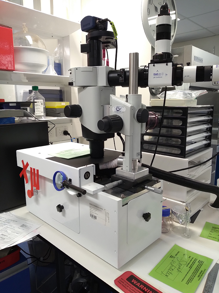 High-resolution episcopic microscopy is a is a technique for visualizing tissue samples, such as mouse embryos, as a large stack of successive 2D images. Detailed description of the HREM technique and examples can be found on the Indigo Scientific website. Consisting of a motorized microtome, fluorescence stereomicroscope and camera, the HREM cuts away a sample at 1-5 micron intervals and photographs the remaining faceplate, section by section. The result is hundreds or thousands of perfectly aligned images, with resolution approaching that of conventional histology, that are available for reassembling in 3D. Datasets captured on the HREM can then be used to generate 3D images through assistance from the Microscopy and Imaging Facility and our available reconstruction software packages.