Analysis Workstations and Software
SBMS Voltage Supercomputer
20,000 core processors located on multiple GPU Volta cards accessible through the Microsoft Remote Desktop application. Advanced image rendering of large data sets and data storage on 200 terabytes of disk space. Access requires a formal induction!
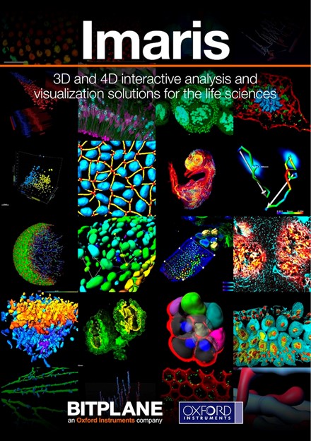 Imaris
Available on SBMS Voltage Supercomputer and stand-alone Computer Workstations located in Otto and Skerman buildings.
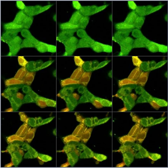
Imaris is Biplane’s core software module that delivers all the necessary functionality for data visualization, analysis, segmentation and interpretation of 3D and 4D microscopy datasets. Combining speed, precision and ease-of-use, Imaris provides a complete set of features for working with three- and four-dimensional multi-channel images of any size, from a few megabytes to multiple gigabytes in size. Conveniently load, process and visualize images acquired from almost any confocal and wide field microscope to gain new and groundbreaking insight from your image data.
Visit the Scitech website for more information.
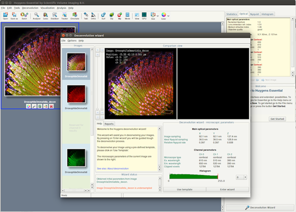 Huygens Deconvolution
Available on SBMS Voltage Supercomputer
Deconvolution is a mathematical operation used in image restoration to recover an object from an image that is degraded by blurring and noise. In fluorescence microscopy the blurring is largely due to diffraction limited imaging by the instrument.
The Huygens Software includes many image processing options, for brightfield and all types of fluorescence Microscope images. With Huygens it is possible to perform image deconvolution and restoration, interactive analysis and volume visualization in 2D, 3D, multi-channel and time.
Beautiful and true images are easily made with a host of visualization tools in Huygens like the MovieMaker, Ortho and Twin slicers, and the Volume, MIP, and Surface renderers.
Visit the SVI website for more information.
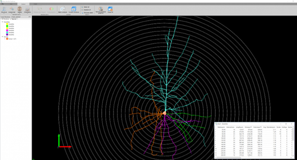 Neuolucida Explorer and 360
Available on SBMS Voltage Supercomputer
Neurolucida Explorer is the analytical software companion for Neurolucida and Neurolucida 360 and is designed to perform extensive morphometric analysis on neuron reconstructions, serial section reconstructions and brain maps that were created by Neurolucida and Neurolucida 360. It also allows you to visualize and analyze data in a 3D environment while exploring the quantitative measurements. Neurolucida Explorer is highly interactive, allowing researchers to explore their data models and see quantitative metrics generated directly from the models.
Neurolucida Explorer is the most comprehensive solution available for neuroanatomical morphometric analysis. It contains dozens of analyses that allow you analyze thousands of parameters. With Neurolucida Explorer, you can customize many of the analyses to address your specific research questions. You can also easily export the results to most statistical software and spreadsheet software for analysis of populations.
Visit the MBF Bioscience website for more information.
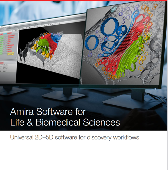 Amira
Amira
Thermo Scientific Amira Software is a powerful, comprehensive, and versatile software solution for visualizing, analyzing and understanding life science and biomedical research images from many image modalities, including Optical and Electron Microscopy, CT, MRI and other imaging modalities.
 Zeiss ZEN Blue
More than 180 image processing tools help you transform and manage your data. Simply search the keyword of your intended method, e.g., kymograph or deconvolution, ZEN will lead you straight to it. ZEN will read the metadata of the input image, then display only the logical processing steps, and optimize the default parameters automatically. You can even process images from other platforms using third-party import tools. With a dedicated workspace, you can also batch process multiple images with ease for quantitative and unbiased results.