HuCS-ID Lab: Data resources
The HuCS-ID lab has developed several specialized data resources that the research group and students within the group can leverage for their research studies/degrees in craniofacial or skeletal identification. These resources additionally facilitate advanced subject matter workshops to industry / government, per consultancies arranged through UQ’s Research Consultancy, Tenders and Contracts Management Office (CCMO).
The resources include two sets of ultra-resolution medical images (MR and CT) for studying craniofacial anatomy and a specialized library of skeletons with matching pre-skeletisation radiographs (as pertinent to identification).
AP Stephan also curates the Collaborative Facial Soft Tissue Depth Data Store (at CRANIOFACIALidentification.com), which is an open-access, free, and online data resource for all craniofacial identification practitioners to use.
Further details on these data resources can be found below.
HuCS-ID MR Head Dataset (Living Persons)
This resource comprises multiple 3T MRI scans of 184 healthy living subjects. Each individual comes with 2x or 3x scans including: T1-weighted MRI, T2-weighted MRI and PETRA ultrashort echo time. The scan resolutions are ultra-high (0.65 mm slice thickness) to provide very clear/exquisite anatomical detail. The age range of individuals in the sample is 18-74 yrs, with most people under 45 years (mean age = 26 yrs)
All scans are acquired at UQ’s Centre for Advanced Imaging (CAI) using a Siemens Magnetrom Prisma 3T whole body MR.
The final scans for this data resource were acquired in Sep 2025. These scans were collected in collaborative liaison between the SBMS School of Anatomy and the CAI as part of Hona’s PhD work entitled: “Correlations of human facial soft tissue thickness with body mass in sub-adults and adults as revealed by lateral radiographs and MRI”. Participant recruitment and scan supervision was conducted by TWPT Hona.
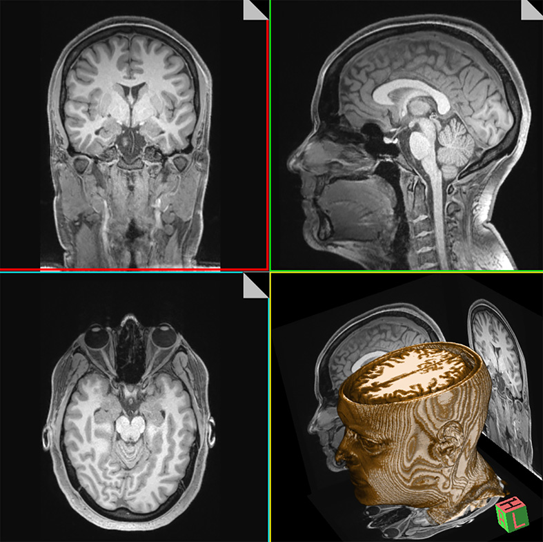
HuCS-ID CT & MR Head Dataset (Decedents)
This resource comprises both CT and MR scans of 92 body donors to UQ’s anatomy program. Five scans represent each individual: one axial CT, one sagittal CT, and three sets of 3T MRIs (T1-weighted MRI, T2-weighted MRI and PETRA ultrashort echo time). The scan resolutions are ultra-high to provide exquisite anatomical detail (0.5 mm slice thickness for each of the CTs and 0.65 mm slice thickness for most MRIs).
This dataset holds the advantage that the CT slice thicknesses are very thin and exist for the full length of the head. Such scans are not attainable for living subjects in the applied clinical setting where the high-resolution parameters cause the scans to exceed acceptable radiation dose limits for living subjects.
All scans are acquired at UQ’s Centre for Advanced Imaging (CAI) using their latest Siemens equipment/software (Biograph Horizon CT scanner and Magnetrom Prisma 3T).
The final scans for this data resource were acquired in Dec 2024. Scan supervision was undertaken by Hona and Stephan (primarily Hona) and in collaborative liaison between the SBMS School of Anatomy and the CAI as part of TWPT Hona’s PhD work entitled: “Correlations of human facial soft tissue thickness with body mass in sub-adults and adults as revealed by lateral radiographs and MRI”.
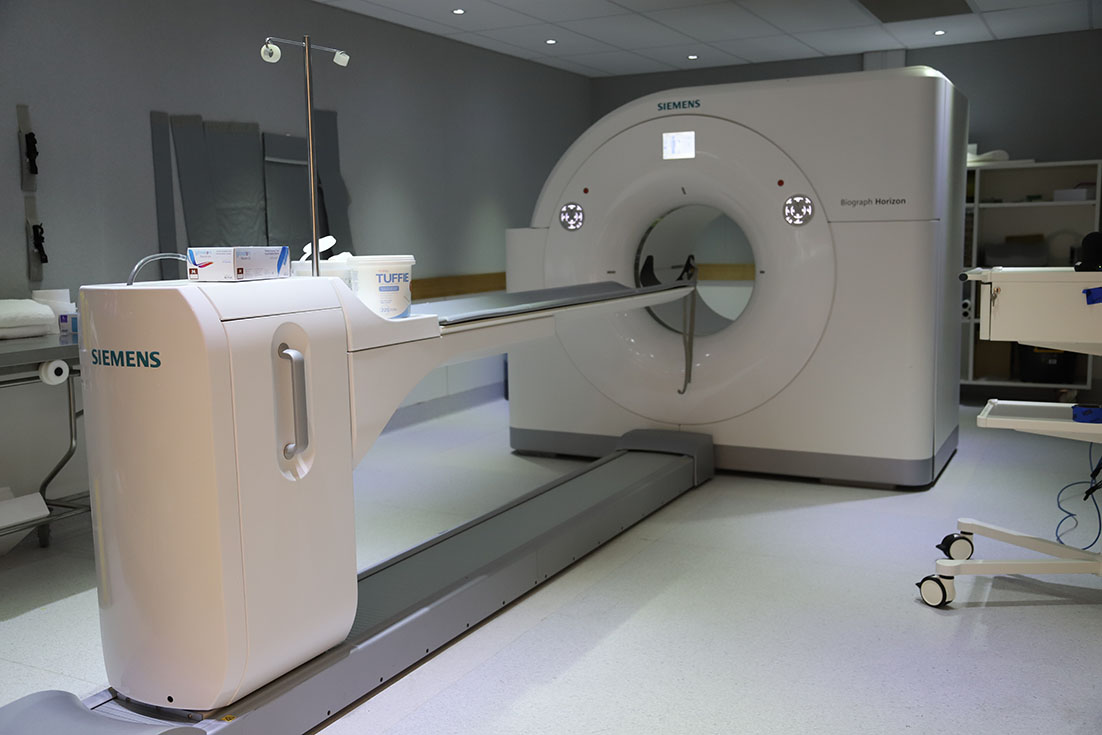
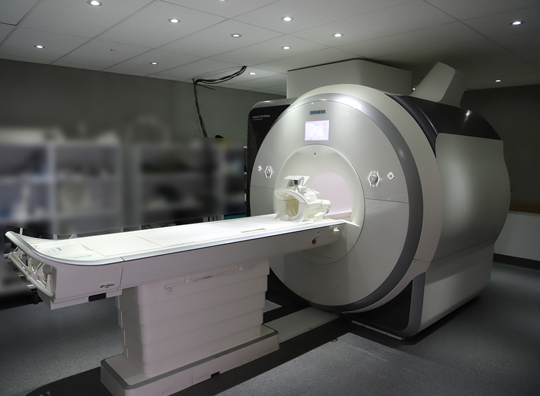
CT and MR machines used for data collection at the CAI.
SBMS Skeletal Collection
This data resource comprises radiographs and skeletons belonging to a subset of Type II (indefinite) body donors to the UQ Body Donor and Anatomy program.
The dry skeletons are 100% traceable to single individuals of origin, and each is accompanied by 8-10 standard radiographs taken prior to skeletisation and all are ethically resourced through the UQ Body Donor Program. These three attributes set this collection apart from other skeletal collections available in the global domain.
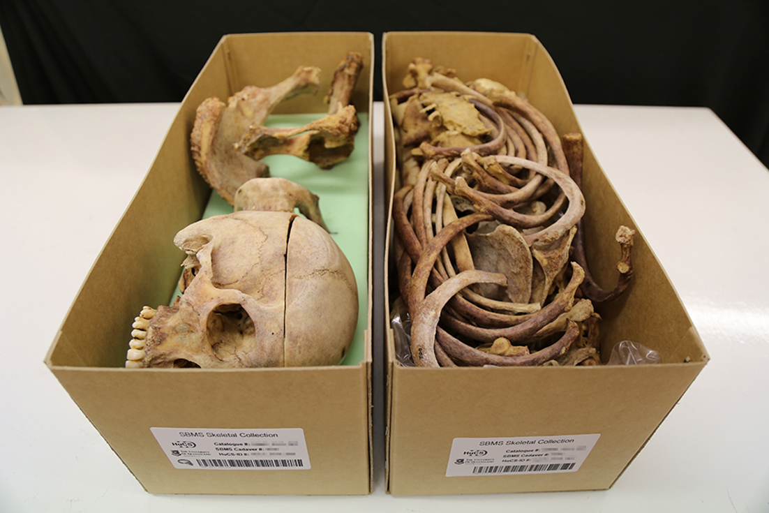
Image reproduced from: Stephan, C. N., Caple, J. M., Veprek, A., Sievwright, E., Kippers, V., Moss, S., & Fisk, W. (2017). Complexities and Remedies of Unknown-provenance Osteology. In N. Pather & G. Strkalj (Eds.), Commemorations and Memorials in Anatomy: Tribute to the Giver (pp. 65-95).
The Collaborative Facial Soft Tissue Depth Data Store (C-Table)
The C-table represents a publicly available and centralised online repository of anonymized raw facial soft tissue depth data for the field of craniofacial identification. It is free to access and separately maintained and updated on a continuing basis by Associate Professor Stephan. Currently, the repository holds data contributed by 20 different research teams (see the C-Table website for details), representing >1,700 individuals collected at up to 25 standardized craniofacial landmarks.