Stephan Group – The Laboratory for Human Craniofacial & Skeletal Identification (HuCS-ID Lab)
The HuCS-ID Lab is a forensic bioanthropology research lab. We use our expertise to improve a wide variety of skeletal identification methods, including those that present some of the toughest challenges in the field - like craniofacial identification. Our goal is to advance the ease, accuracy and reliability of skeletal identification in the medicolegal context. We use a wide variety of advanced methods including medical imaging (MR and CT), 3D scanning, computational analysis, plain film X-ray and dissection to attain our research goals.
Our work crosses both academics and applied science. We hold, for example, a long and valued track-record of work with the USA’s Defense POW/MIA Accounting Agency to assist their noble mission of the fullest possible accounting of missing USA service members.
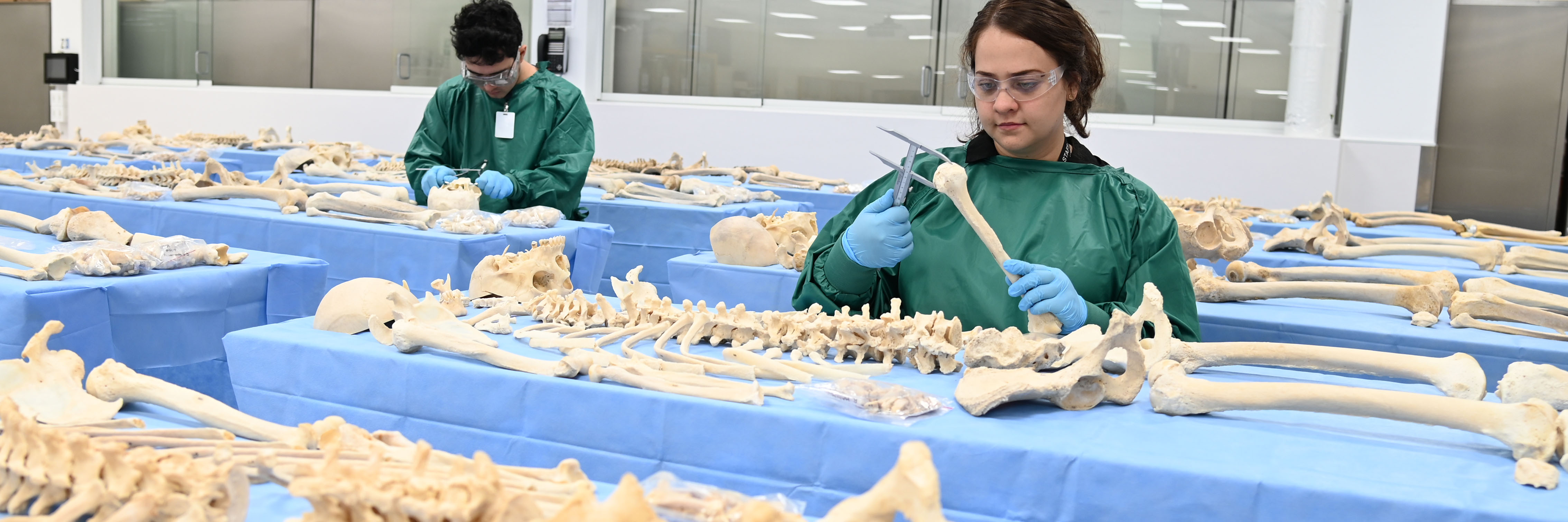
We work very closely with SBMS’s Gross Anatomy Facility (GAF) and this includes management of the GAF’s Skeletisation Program and SBMS Library of Skeletons from indefinite (Type 2) body donors who consent to their body being considered for use in the program. This skeleton library, per its consents, radiographs, traceability and methods, is one of a kind within Australia and globally (see Data resources tab). Donors within the library not only help to teach enrolled UQ students and/or board-certified specialists at advanced professional workshops, they also routinely provide forensic identification advances via research. Recent contributions include radiographic comparison and skeletal unmingling methods in the context of repatriation from past wars and conflicts.
Here is a quick snippet of the type of work we do, as compiled for the American Academy of Forensic Sciences (AAFS) 2024 Meeting in Denver Colorado USA on AAFS|TV:
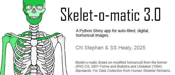
Find out more about the Skelet-o-matic 3.0, the world's first auto-fill digital homunculus produced by some of our team members.
Interests and expertise
Human Identification, Skeletal Identification, Forensic Anthropology, Physical Anthropology, Skeletal Analysis, Osteology, Anatomy, Human variation, Radiographic Comparison, Osteometric Sorting, Mass Graves, Sex estimation, Age estimation, Ancestry estimation, Stature estimation, Trauma, Craniofacial Identification, Skull, Face, Facial approximation, Craniofacial superimposition.
Shape Analysis, Morphometrics, Anthropometry, 3D Scanning, Stereophotogrammetry, Geometric Morphometrics, Elliptical Fourier Analysis.
Medical Imaging, Ultrasound, Radiography, X-Rays, Optics, Photography.
R, Biostatistics, Mechatronics.
Facilities
The HuCS-ID Lab is well-equipped with the usual precision instruments of osteometric boards, callipers and other standard anthropometry devices and we use only internationally recognized manufacturers (such as GPM, Mopec). Instruments are routinely performance checked in-house under an annual quality assurance program and using an array of calibrated test artefacts to ensure measurement validity for scientific studies and casework.
The laboratory holds a broad range of 3D scanning devices to include: Artec Spider, Einscan, Next Engine, and Di3D Stereophotogrammetry. The Di3D equipment, used for instantaneous 3D acquisition of human faces, is operated in a separate dedicated video green-room.
Professional-grade camera equipment is used throughout the laboratory, including Canon 6D and Nikon D780 full-frame DSLRs, mounted on copy stands for detailed skeletal photography.
Being on UQ St Lucia Campus, we are only just down the road (200m) from the UQ Centre for Advanced Imaging (CAI), which offers a large array of medical imaging technology and is where we undertake our CT and MRI scans for research.
The HuCS-ID Lab works closely with the SBMS Gross Anatomy Facility (GAF) and its anatomical resources, so much so, that our forensic anthropology lab is physically embedded in the GAF to ensure seamless workflow under the Transplantation and Anatomy Act, Qld (1979). The UQ School of Anatomy is one of the largest anatomy Schools in Australia and it is the only school of its kind in the country to operate a whole-of-body skeletisation program for eligible indefinite donors.
Introduction
Anthropological instruments
Scan capabilities
The HuCS-ID lab has developed several specialized data resources that the research group and students within the group can leverage for their research studies/degrees in craniofacial or skeletal identification. These resources additionally facilitate advanced subject matter workshops to industry / government, per consultancies arranged through UQ’s Research Consultancy, Tenders and Contracts Management Office (CCMO).
The resources include two sets of ultra-resolution medical images (MR and CT) for studying craniofacial anatomy and a specialized library of skeletons with matching pre-skeletisation radiographs (as pertinent to identification).
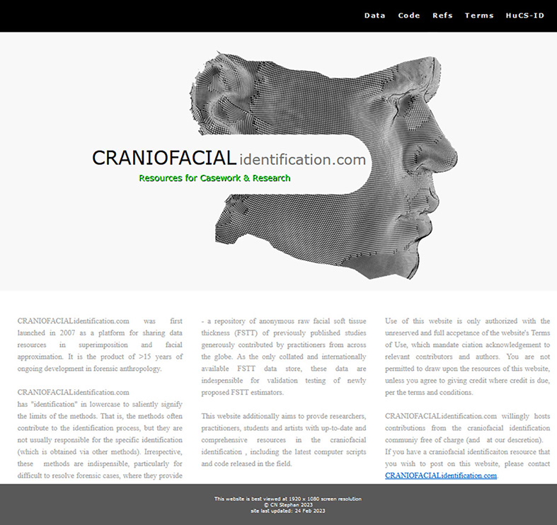
AP Stephan also curates the Collaborative Facial Soft Tissue Depth Data Store (at CRANIOFACIALidentification.com), which is an open-access, free, and online data resource for all craniofacial identification practitioners to use.
Further details on these data resources can be found below.
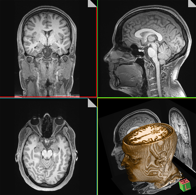
HuCS-ID MR Head Dataset (Living Persons)
This resource comprises multiple 3T MRI scans of 184 healthy living subjects. Each individual comes with 2x or 3x scans including: T1-weighted MRI, T2-weighted MRI and PETRA ultrashort echo time. The scan resolutions are ultra-high (0.65 mm slice thickness) to provide very clear/exquisite anatomical detail. The age range of individuals in the sample is 18-74 yrs, with most people under 45 years (mean age = 26 yrs)
All scans are acquired at UQ’s Centre for Advanced Imaging (CAI) using a Siemens Magnetrom Prisma 3T whole body MR.
The final scans for this data resource were acquired in Sep 2025. These scans were collected in collaborative liaison between the SBMS School of Anatomy and the CAI as part of Hona’s PhD work entitled: “Correlations of human facial soft tissue thickness with body mass in sub-adults and adults as revealed by lateral radiographs and MRI”. Participant recruitment and scan supervision was conducted by TWPT Hona.
HuCS-ID CT & MR Head Dataset (Decedents)
This resource comprises both CT and MR scans of 92 body donors to UQ’s anatomy program. Five scans represent each individual: one axial CT, one sagittal CT, and three sets of 3T MRIs (T1-weighted MRI, T2-weighted MRI and PETRA ultrashort echo time). The scan resolutions are ultra-high to provide exquisite anatomical detail (0.5 mm slice thickness for each of the CTs and 0.65 mm slice thickness for most MRIs).
This dataset holds the advantage that the CT slice thicknesses are very thin and exist for the full length of the head. Such scans are not attainable for living subjects in the applied clinical setting where the high-resolution parameters cause the scans to exceed acceptable radiation dose limits for living subjects.
All scans are acquired at UQ’s Centre for Advanced Imaging (CAI) using their latest Siemens equipment/software (Biograph Horizon CT scanner and Magnetrom Prisma 3T).
The final scans for this data resource were acquired in Dec 2024. Scan supervision was undertaken by Hona and Stephan (primarily Hona) and in collaborative liaison between the SBMS School of Anatomy and the CAI as part of TWPT Hona’s PhD work entitled: “Correlations of human facial soft tissue thickness with body mass in sub-adults and adults as revealed by lateral radiographs and MRI”.
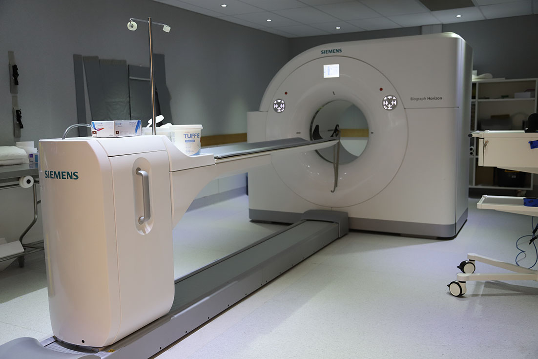
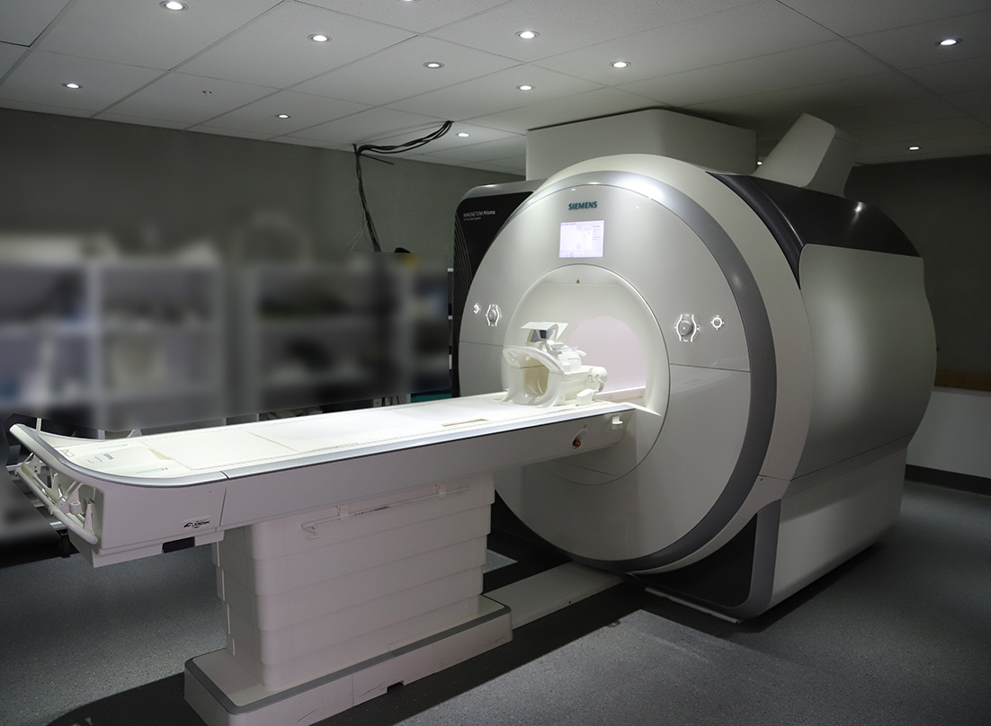
CT and MR machines used for data collection at the CAI.
SBMS Skeletal Collection
This data resource comprises radiographs and skeletons belonging to a subset of Type II (indefinite) body donors to the UQ Body Donor and Anatomy program.
The dry skeletons are 100% traceable to single individuals of origin, and each is accompanied by 8-10 standard radiographs taken prior to skeletisation and all are ethically resourced through the UQ Body Donor Program. These three attributes set this collection apart from other skeletal collections available in the global domain.
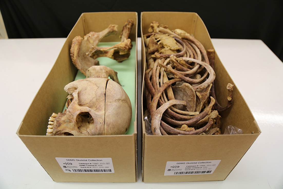
Image reproduced from: Stephan, C. N., Caple, J. M., Veprek, A., Sievwright, E., Kippers, V., Moss, S., & Fisk, W. (2017). Complexities and Remedies of Unknown-provenance Osteology. In N. Pather & G. Strkalj (Eds.), Commemorations and Memorials in Anatomy: Tribute to the Giver (pp. 65-95).
The Collaborative Facial Soft Tissue Depth Data Store (C-Table)
The C-table represents a publicly available and centralised online repository of anonymized raw facial soft tissue depth data for the field of craniofacial identification. It is free to access and separately maintained and updated on a continuing basis by Associate Professor Stephan. Currently, the repository holds data contributed by 20 different research teams (see the C-Table website for details), representing >1,700 individuals collected at up to 25 standardized craniofacial landmarks.
The HuCS-ID Lab publishes in leading forensic science and biological anthropology journals.
Examples include:
The International Journal of Legal Medicine, The American Journal of Biological Anthropology, The Journal of Forensic Sciences, Forensic Science International, Science & Justice, and Forensic Imaging.
Group Head
Students


GAF Staff Collaborators

Spotlight on HuCS-ID student alumni
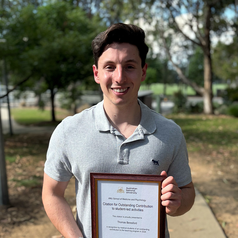 Tom Beresford
Tom graduated Honours with a First Class in the lab in 2022 for work entitled “The Accuracy of 3D Scanners for Recording Human Skeletal Anatomy”. He presented this work in-person at a special session for the Humanitarian and Human Rights Resource Center (HHRRC) in Orlando USA during the 75th Annual Meeting of the American Academy of Forensic Sciences, 2023. In 2024, Tom began a Doctor of Medicine and Surgery (MChD) at ANU, attaining grades within the top 3% of his class. Also in 2024, he received a Faculty Citation for Outstanding Contribution to Student Led Activities (first-rate, unpaid, anatomy tutoring of his colleagues also enrolled in MChD). Tom’s Honours work is presently under consideration for publication with Forensic Science International.
 Dr Hayley Fancourt
Hayley graduated with a First Class Honours in Science (2017). Hayley published one paper during her Honours year (FSI) and another one following degree completion (JFS). Hayley attained both the highest score for her final research presentation and won the Michael F. Hickey Memorial Honours Prize (highest overall Honours grade for the SBMS in that year). Hayley recently graduated Medicine from the Australian National University, where she also worked part time as an Anatomy Technical Officer.
 Dr Jodi Caple
Obtained her PhD in 2018, graduating with 6 paper outputs from the PhD and received one of three Dean’s Awards for an Outstanding Thesis. Dr Caple now works as a Forensic Anthropologist & Postdoctoral Research Associate with SNA International at the Defense POW/MIA Accounting Agency, HI, USA.
Lab alumni
PhD
- Telena Hona: Correlations of human facial soft tissue thickness with body mass in sub-adults and adults as revealed by lateral radiographs and MRI
- Sean Healy: Craniofacial superimposition: Can machine learning improve focus distance estimation from real-world facial photographs
- Jeffrey Lynch: Computerized methods for de-commingling mixed human skeleton assemblages: New Automated Approaches Using Centroid Banding
- Jodi Caple: Fourier Analysis of Lateral Cranial Profiles for Predicting Sex and Ancestry in Forensic Anthropology
Honours
- Rebecca Menzie, Optimising anatomy practical exams to benchmark student understanding and maximise the student experience (active 2025-26)
- Aariz Khan, Do truncated spinous processes impact radiographic shadows for identification? (active 2025-26)
- Judy Magirin, Variation of Sphenoid Sinuses from Segmented CT and MR Multislice Reconstructions (active 2025)
- Yong Wei Xue, Nose Estimation from the Skull Using Ultra-high Resolution CT Scans (active 2025)
- Robert Humphrey: An anatomical CT investigation of the eyeball relationships to the bony orbit
- Lachlan McInerney: The anatomical accuracy of mouth width estimation methods from the skull: Validation tests using 3D medical scans
- Jithya Joshy: A craniometric dataset to help detect South Asian skulls and filter their intake into Anatomy Teaching Schools
- Suhail Askarzai: Does Phalangeal Morphology Encode Side, Digit and Row as Relevant for Legal Medicine?
- Sean Healy: Improving identification for forensic craniofacial superimposition: The accuracy of subject-to-camera distance estimation from human face anatomy recorded in frontal and profile images.
- Tom Beresford: How good are 3D optical scanners and photogrammetry for scientific analysis of human bone anatomy?
- Te Wai Pounamu Telena Hona: Accuracies of Radiographic Comparison for Skeletal Identification
- Brianna Armstrong: Testing the accuracy of subject-to-camera distance estimation in craniofacial superimposition using palpebral fissure length
- Madeline Ashton: DNA tracking for embalmed specimens
- Chelsea Olditch: Radiographic Comparison for Human Identification
- Brandon Meikle: Patterns of Facial Soft Tissue Thicknesses as Measured in Large Samples by Ultrasound
- Jennifer Atkins: The utility of infracranial methods for sex and age estimation of contemporary Australians
- Hayley Fancourt: Pairwise matching of commingled long bones: a computerized approach using 3D elliptical Fourier analysis
- Redmond Lopez: Sex and Ancestry Variation of the Extracranial Space
- Ashleigh Yeap: Characterisation of Facial Muscle Morphology from Bony Attachments
- Emma Sievwright: Do Craniometrics Correlate with Facial Soft Tissue Thicknesses in a Living Adult Australian Sample
- Lachlan Munn: Posture and Expression Effects on Face Typology as Mapped by 3D Stereophotogrammetry for Forensic Face Identification
- Rory Preisler: The effect of Posture on Facial Soft Tissue Depths, Shorths and Shormaxes – Using B-mode Ultrasound
- Jodi Caple: Facial Soft Tissue Thicknesses of Australian Juveniles: Analysis of the Computed Tomography Tangent Method
November 2025
- Last week, Ullrich and Stephan’s 2016 article Mikhail Mikhaylovich Gerasimov's Authentic Approach to Plastic Facial Reconstruction was the third-most-read research item from SBMS in ResearchGate.
October 2025
- The latest 2025 composite score (v8 released Oct) ranks Carl 36 most cited author globally in Legal & Forensic Medicine (1st Science Metrix Category), up 28 places from 2019.
This places Carl in the top 0.25% of 15,009 forensic scientists world-wide who hold more than 5 paper publications and a top ranked Science-Metrix category of Legal & Forensic Medicine.
Globally, from all fields, disciplines and scientists Carl ranks 109,028 (up 25,378 places from 5 years ago).
Reference: Baas, Boyack & Ioannidis (2025), Data for "Updated science-wide author databases of standardized citation indicators", Elsevier Data Repository, v8. - Congratulations to Judy and Yong for completing final Honours Reports and Seminars this week (22-Oct)! Outstanding efforts and with improved casework relevant methods!
- Ullrich and Stephan’s (2016) article was the second-most-read research item from SBMS in ResearchGate last week (13-17 Oct). A top three placing is becoming a regular occurrence!
- The HuCS-ID Lab is looking forward to the special forensic anthropology session to be held at the Australasian Society for Human Biology Meeting this year in Auckland later this year (Dec). We will be there and hope to see you too!
September 2025
- Carl Stephan delivered two invited scientific talks at DPAA’s 2025 Scientific Summit on the Repatriation of Persons from Prior Military Operations / Wars (hosted by LABANOF) at the State University of Milan, Italy.
One presentation concerned the strategy and formulation of standardized chest X-ray comparison methods (with multiple quality assurance layers) at the DPAA.
The second seminar regarded updated craniofacial superimposition methods that were pivotal in resolving a long standing DPAA Korean War case, for which DNA and dental records could not offer conclusive answers. - For the last week of this month, Ullrich and Stephan’s (2016) article was the second-most-read research item from SBMS in ResearchGate.
August 2025
One of Carl’s facial reconstruction papers featured as the most read paper from SBMS on research gate last week (18–24 Aug). See Mikhail Mikhaylovich Gerasimov's Authentic Approach to Plastic Facial Reconstruction.
May 2025
For two weeks of last month Ullrich and Stephan’s 2016 article – Mikhail Mikhaylovich Gerasimov's Authentic Approach to Plastic Facial Reconstruction – was the third most read paper from SBMS in research gate.
April 2025
- Carl and Sean released the world’s first digital auto-fill homunculus capability, Skelet-o-matic 3.0! This free python script (licensed under GPL version 2) enables skeletal homunculus files (.png, .jpg, .tiff) to be generated in seconds without tedious by-hand colouring of skeleton outline charts. It is available for all to use to make skeletal homunculi more user-friendly. Watch a video demo.
January 2025
- In Jan we host the 9th Masterclass for DPAA analysts in Chest Radiograph Comparison onsite at UQ.
We are all very much looking forward to welcoming the new DPAA analyst team to Brisbane and UQ for the training!
December 2024
- Our first CT & MR craniofacial dataset of decedents has reached maturity with workable research sample size of 91 participants! See the Data Resources tab for more info.
- We’ve received ethics approval to proceed with our next craniofacial MR dataset on living individuals (0.65mm slices 3 protocols, and 0.4mm slices 1 protocol)! See the Data Resources tab for more info.
- Congratulations to Rob Humphrey who, after completing Honours in our Lab analyzing CT scans, was just awarded a place to study Medicine at Griffith University! Well done, Rob!
November 2024
- A big congrats go to Lachie and Rob for successfully completing their Honours research studies this month with outstanding results! Amazing efforts made and with publication worthy results!
October 2024
- This month the HDR members in the lab and the team leader, all attended the 20th Meeting of the International Association for Craniofacial Identification in Granada, Spain.
It was a blast to catch up with colleagues and discuss the latest science! In total the HuCS-ID lab made 5 scientific presentations and delivered one workshop on R.
Congrats go to, Ronn Taylor, a long-standing Australian icon of facial approximation, mentor and colleague, who received a IACI Lifetime Achievement Award at the meeting!
May 2024
- This month we completed our 8th Masterclass for DPAA analysts in Chest Radiograph Comparison onsite at UQ.
April 2024
- Jithya Joshy has been announced as the winner of the Michael F. Hickey Prize for the highest overall SBMS Honours research degree mark, awarded in Anatomy & Physiology subdisciplines for 2023 – congratulations Jithya!
February 2024
- Pioneering work of the HuCS-ID Lab and UQ Anatomy Lab regarding Medical Teaching Skeletons, first undertaken in 2017 and leading ethical interventions towards these remains in the anatomy space, receives attention in the lastest issue of Nature Communications 2024.
For additional information on nuances of these medical teaching skeletons see Stephan et al. 2019 Skeletal Evidence of Sharp–Force Disarticulation and Tissue Flensing in 54 Cases Exhibiting Approximately 4,200 Bone Strike Injuries.
Chest radiograph comparisons continue to help resolve the identities of unaccounted-for US Soldiers from the Korean War at the USA Defense POW/MIA Accounting Agency (DPAA).
In the first 7 weeks of this year, three identifications from the Korean War have been made by the DPAA and all three included Chest Radiograph Comparisons as a line of identification evidence.
For further information see DPAA News Releases (also includes links to releases for prior accounting years).
- The American Academy of Forensic Sciences (AAFS) features the HuCS-ID Lab, Dr John Byrd (DPAA Laboratory Director) and Michael Streed (The SketchCop) in the AAFS |TV Thought Leadership Video “Forensic Anthropology at the Front Lines of Repatriation & Skeletal Identification”. Watch here.
January 2024
Websedge begins filming at the HuCS-ID Lab for the 2024 AAFS|TV Thought Leadership Film Series – Look out for the short films at the American Academy of Forensic Sciences Meeting in Denver, Feb 2024!
The HuCS-ID Lab takes video superimposition to the photographics literature for the first time, reframing methods in terms of critical photographic parameters – see our latest publication in the Journal of Imaging.
December 2023
- Congratulations to Sean Healy on winning 2023’s SBMS Student Research Excellence Award at the recently held SBMS Awards Night!
HuCS-ID Lab students present 3 papers at this year’s Australasian Society of Human Biology (ASHB) Conference in Brisbane – well done Sean, Telena and Jithya.
Congratulations to Jithya Joshy on obtaining the runners-up prize for Best Student Presentation at ASHB, Brisbane, on her talk concerning medical teaching skeletons!
November 2023
Congratulations to Telena on her recent award of a 2023 American Academy of Forensic Sciences (AAFS) Student Travel Grant to attend and present at the next annual AAFS meeting in Denver 2024! We send acknowledgements of thanks to AAFS for their generous support of international students (and their long-haul flights from remote locations)!!
Congratulations to Tom Beresford on his acceptance to the ANU Medical Program for 2024! Outstanding achievement Tom!
October 2023
Congratulations Jithya on obtaining the second highest mark in the SBMS Honours cohort for your final report seminar! Talk title: “A craniometric dataset to help detect South Asian skulls and filter their intake into Anatomy Schools”
The latest 2023 composite (c) score based on Scopus data has just been released placing Carl’s work in the top 0.3% of all forensic scientists who hold: a) more than 5 paper publications; and b) a top ranked Science-Metrix category of Legal & Forensic Medicine. USA rank = 14th, worldwide rank = 77th when Legal & Forensic Medicine is either the first OR second Science-Metrix category. Reference: Ioannidis, John P.A. (2023), “October 2023 data-update for "Updated science-wide author databases of standardized citation indicators"”, Elsevier Data Repository, V6, doi: 10.17632/btchxktzyw.6
September 2023
- PhD students in the Lab strike a record with 3 papers accepted in IJLM within one month!!! Way to go Sean and Telena!
July 2023
- The HuCS-ID Lab hosted the 7th Masterclass Training in Chest Radiograph Comparison for DPAA analysts onsite at UQ (St Lucia and Herston Campuses).
April 2023
- Congratulations to Telena Hona on winning the American Association of Biological Anthropologists’ (AABA) Prize for Outstanding Student Presentation: Anatomy in Anthropology, Reno, USA for her podium research talk: Understanding Childhood Growth—A Pilot Study of Facial Soft Tissue Thicknesses and Body Mass Relationships in Children Aged 5-17 years!
March 2023
- HuCS-ID Lab begins new work establishing the first of two new hallmark head datasets comprised of medical images that will provide unrivalled research resources in craniofacial ID for the future.
This first dataset, the HuCS-ID CT & MRI Head Dataset (Decedents), comprises 5x 3D scans of 70 decedent heads (2x different CT protocols and 3x different 3T MR protocols all at the super-resolution of 0.5-0.65mm slice thickness!).
The second dataset, HuCS-ID MR Head Dataset (Living Persons), will be initiated later this year (3x different 3T MR protocols all at the super-resolution of 0.5-0.65mm slice thickness).
These data provide outstanding opportunities for new PhD students. Sign up now!
February 2023
- Congratulations to Telena Hona on her receipt of a 2023 Pollitzer Student Travel Grant to attend the next American Association of Biological Anthropologists (AABA) Meeting in Reno, USA!
- Fancourt et al. (2021) paper on commingling has been recognized by Wiley as a JFS Top Cited paper for 1 Jan 2021-15 Dec 2022 (Clarivate Analytics)
January 2023
The HuCS-ID Lab is celebrating its 10th year at UQ and in SBMS this month.
The HuCS-ID Lab hosted the 6th CXR Masterclass Training for DPAA analysts onsite UQ at St Lucia and Herston Campuses. Jump to DPAA’s Linkedin post on the training.
December 2022
- UQ Faculty of Medicine's UQmedicine magazine profiles Associate Professor Carl Stephan
October 2022
- The HuCS-ID Lab is now accepting Honours Research applications for 2023.
A number of forensic anthropology and anatomy projects are on offer. Contact the Lab Head, Assoc Prof Stephan, for more information.
- HuCS-ID Lab, with Telena Hona as lead author, publishes in Forensic Imaging—the first paper in the journal’s history to receive, from all three expert reviewers, “a publish with no corrections”!
Notified by Dr Thomas Ruder, Associate Editor, Forensic Imaging
September 2022
The Journal of Forensic Sciences Recognizes a 2017 HuCS-ID Lab paper as outstanding by Inclusion in their National Forensic Science Week Virtual Special Issue!
DPAA Lab Director, Dr John Byrd, outlines contributions of Chest Radiograph comparison to Defense POW/MIA Accounting Agency Mission on Youtube. Watch it now.
- DPAA showcases a video sequence of the Clavicle Matching Program (CMP) on their Facebook #ForensicFriday pages.
July 2022
- Congratulations to Sean Healy (Honours student) for his win of a Best Student Presentation at the 19th Annual Meeting of the International Association for Craniofacial Identification in Liverpool UK.
- AP Stephan’s Best Scientific Abstract Award at the 19th International Association of Craniofacial Identification Meeting (2022) was received by Seppe Goovaerts (Belgium) and colleagues for the abstract “Multivariate GWAS identifies 30 independent genetic loci involved in normal-range 3D cranial vault shape”.
May 2022
- The HuCS-ID Lab hosted the 5th CXR Masterclass Training for DPAA analysts onsite UQ at St Lucia and Herston Campuses.
August 2018
- The HuCS-ID Lab welcomes the International Association of Craniofacial Identification (IACI) to Brisbane for their 30th anniversary meeting, with multiple team members contributing to a FSI special issue: Latest Progress in Craniofacial Identification: 17th Biennial Meeting of the International Association of Craniofacial Identification (IACI), Brisbane, 15-19 July 2017